Structure and Function
The eye transmits visual stimuli to the brain for interpretation and, in doing so, functions as the organ of vision. The eyeball is located in the eye orbit, a round, bony hollow formed by several different bones of the skull. In the orbit, the eye is surrounded by a cushion of fat. The bony orbit and fat cushion protect the eyeball. To perform a thorough assessment of the eye, you need a good understanding of the external structures of the eye, the internal structures of the eye, the visual fields and pathways, and the visual reflexes.
transmit:to send an electronic signal, radio or television broadcast, etc. 传送;输送;发射;播送
stimuli:something that produces a reaction in a human, an animal or a plant (使生物产生反应的)刺激,刺激物
orbit:a curved path followed by a planet or an object as it moves around another planet, star, moon, etc. (天体等运行的)轨道
hollow:having a hole or empty space inside 中空的;空心的;(of parts of the face 面部) sinking deeply into the face 凹陷的
cushion:a layer of sth between two surfaces that keeps them apart (隔离两个表面的)垫;something that protects you against sth unpleasant that might happen 起保护(或缓冲)作用的事物
fat:a white or yellow substance in the bodies of animals and humans, stored under the skin 脂肪;肥肉
the visual reflexes:视觉反射
External Structures of the Eye
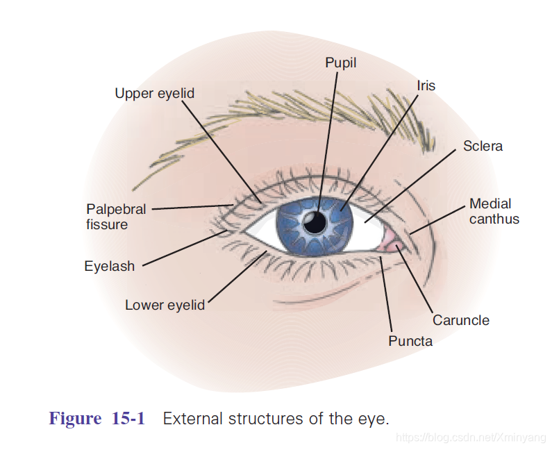
The eyelids (upper and lower) are two movable structures composed of skin and two types of muscle: striated and smooth. Their purpose is to protect the eye from foreign bodies and limit the amount of light entering the eye. In addition, they serve to distribute tears that lubricate the surface of the eye (Fig. 15-1). The upper eyelid is larger, more mobile, and contains tarsal plates made up of connective tissue. These plates contain the meibomian glands, which secrete an oily substance that lubricates the eyelid.
eyelid= either of the pieces of skin above and below the eye that cover it when you blink or close the eye 眼睑;眼皮
striate=有条纹的,有细槽的;
lubricate= to put a lubricant on sth such as the parts of a machine, to help them move smoothly 给…上润滑油;上油于
meibomian glands=睑板腺
secrete= (of part of the body or a plant 身体或植物器官) to produce a liquid substance 分泌
The eyelids join at two points: the lateral (outer) canthus and medial (inner) canthus. The medial canthus contains the puncta, two small openings that allow drainage of tears into the
lacrimal system, and the caruncle, a small, fleshy mass that contains sebaceous glands. The white space between open eyelids is called the palpebral fissure. When closed, the eyelids
should touch. When open, the upper lid position should be between the upper margin of the iris and the upper margin of the pupil. The lower lid should rest on the lower border of the iris. No sclera should be seen above or below the limbus (the point where the sclera meets the cornea).
canthus= ['kænθəs] 眼睛的两端,眼角,眦;
puncta=点; 色斑; 凹陷; 尖;
drainage= the process by which water or liquid waste is drained from an area 排水;放水
lacrimal=泪腺的
caruncle=泪器
sebaceous= (biology 生) producing a substance like oil in the body 分泌脂质的;皮脂腺的
palpebral fissure= [ˈpælpibrəl ˈfɪʃə] 睑裂
iris= the round coloured part that surrounds the pupil of your eye 虹膜
pupil= the small round black area at the centre of the eye 瞳孔;眸子;瞳人
Eyelashes are projections of stiff hair curving outward along the margins of the eyelids that filter dust and dirt from air entering the eye.
The conjunctiva is a thin, transparent, continuous membrane that is divided into two portions: a palpebral and a bulbar portion. The palpebral conjunctiva lines the inside of the eyelids, and the bulbar conjunctiva covers most of the anterior eye, merging with the cornea at the limbus. The point at which the palpebral and bulbar conjunctivae meet creates a folded recess that allows movement of the eyeball. This transparent membrane allows for inspection of underlying tissue and serves to protect the eye from foreign bodies.
Eyelash= one of the hairs growing on the edge of the eyelids 睫;睫毛
stiff= firm and difficult to bend or move 不易弯曲(或活动)的;硬的;挺的
conjunctiva=(眼球)结膜

The lacrimal apparatus consists of glands and ducts that serve to lubricate the eye (Fig. 15-2). The lacrimal gland, located in the upper outer corner of the orbital cavity just above
the eye, produces tears. As the lid blinks, tears wash across the eye then drain into the puncta, which are visible on the upper and lower lids at the inner canthus. Tears empty into the
lacrimal canals and are then channeled into the nasolacrimal sac through the nasolacrimal duct. They drain into the nasal meatus.
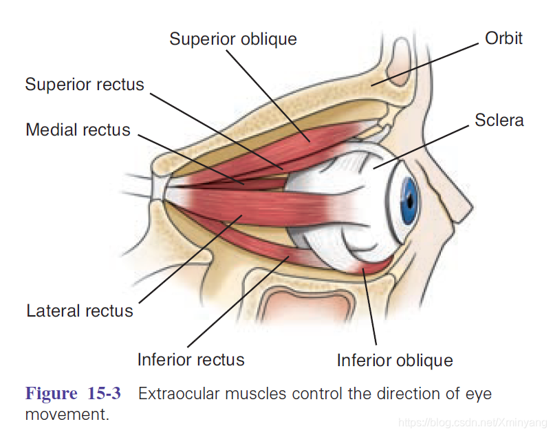
The extraocular muscles are the six muscles attached to the outer surface of each eyeball (Fig. 15-3). These muscles control six different directions of eye movement. Four rectus muscles
are responsible for straight movement, and two oblique muscles are responsible for diagonal movement. Each muscle coordinates with a muscle in the opposite eye. This allows for
parallel movement of the eyes and thus the binocular vision characteristic of humans. Innervation for these muscles is supplied by three cranial nerves: the oculomotor (III) trochlear
(IV), and abducens (VI).
Clinical Tip:Because of this sensory property, contact with a wisp of cotton stimulates a blink in both eyes known as the corneal reflex. This reflex is supported by the trigeminal nerve, which carries the afferent sensation into the brain, and the facial nerve, which carries the efferent message that stimulates the blink.
The middle layer contains both an anterior portion, which includes the iris and the ciliary body, and a posterior layer, which includes the choroid. The ciliary body consists of muscle tissue that controls the thickness of the lens, which must be adapted to focus on objects near and far away.
The iris is a circular disc of muscle containing pigments that determine eye color. The central aperture of the iris is called the pupil. Muscles in the iris adjust to control the pupil’s size, which controls the amount of light entering the eye. The muscle fibers of the iris also decrease the size of the pupil to accommodate for near vision and dilate the pupil when far vision is needed.
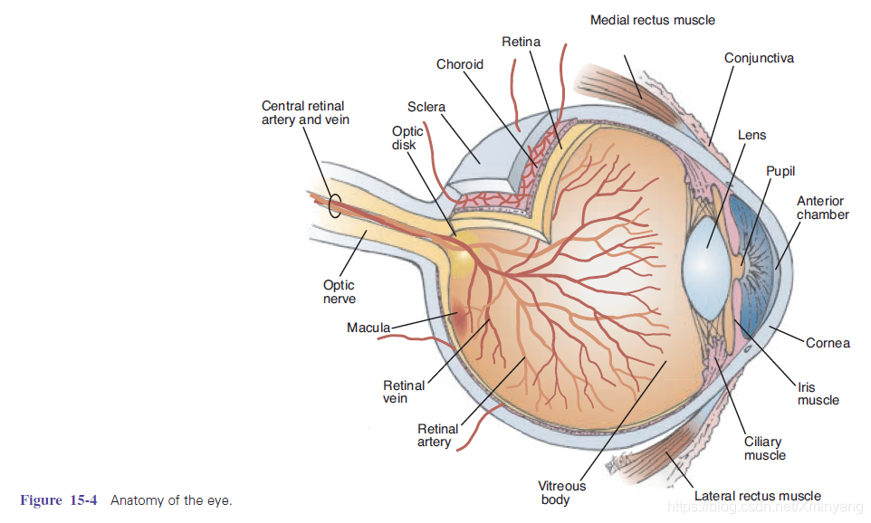
The lens is a biconvex, transparent, avascular, encapsulated structure located immediately posterior to the iris. Suspensory ligaments attached to the ciliary body support the position of the lens. The lens functions to refract (bend) light rays onto the retina. Adjustments must be made in refraction depending on the distance of the object being viewed. Refractive ability of the lens can be changed by a change in shape of the lens (which is controlled by the ciliary body). The lens bulges to focus on close objects and flattens to focus on far objects.
The chorioid layer contains the vascularity necessary to provide nourishment to the inner aspect of the eye and prevents light from reflecting internally. Anteriorly, it is continuous with
the ciliary body and the iris.
The innermost layer, the retina, extends only to the ciliary body anteriorly. It receives visual stimuli and sends it to the brain. The retina consists of numerous layers of nerve cells,
including the cells commonly called rods and cones. These specialized nerve cells are often referred to as “photoreceptors” because they are responsive to light. The rods are highly
sensitive to light, regulate black and white vision, and function in dim light. The cones function in bright light and are sensitive to color.
The optic disc is a cream-colored, circular area located on the retina toward the medial or nasal side of the eye. It is where the optic nerve enters the eyeball. The optic disc can be
seen with the use of an ophthalmoscope and is normally round or oval in shape, with distinct margins. A smaller circular area that appears slightly depressed is referred to as the physiologic cup. This area is approximately one-third the size of the entire optic disc and appears somewhat lighter/whiter than the disc borders.
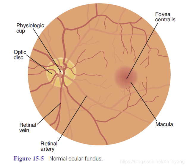
The retinal vessels can be readily viewed with the aid of an ophthalmoscope. Four sets of arterioles and venules travel through the optic disc, bifurcate, and extend to the periphery of
the fundus. Vessels are dark red and grow progressively narrower as they extend out to the peripheral areas. Arterioles carry oxygenated blood and appear brighter red and narrower
than the veins. The general background, or fundus (Fig. 15-5), varies in color, depending on skin color. A retinal depression known as the fovea centralis is located adjacent to the optic
disc in the temporal section of the fundus. This area is surrounded by the macula, which appears darker than the rest of the fundus. The fovea centralis and macular area are highly
concentrated with cones and form the area of highest visual resolution and color vision.
The eyeball contains several chambers that serve to maintain structure, protect against injury, and transmit light rays. The anterior chamber is located between the cornea and iris, and
the posterior chamber is the area between the iris and the lens. These chambers are filled with aqueous humor, a clear liquid substance produced by the ciliary body. Aqueous humor helps to cleanse and nourish the cornea and lens as well as maintain intraocular pressure. The aqueous humor filters out of the eye from the posterior to the anterior chamber then into the canal of Schlemm through a filtering site called the trabecular meshwork. Another chamber, the vitreous chamber, is located in the area behind the lens to the retina. It is the largest
of the chambers and is filled with a vitreous humor that’s clear and gelatinous.
VISION:Visual Fields and Visual Pathways
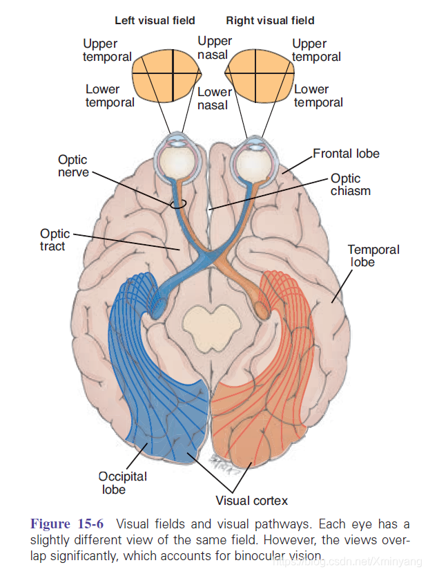
A visual field refers to what a person sees with one eye. The visual field of each eye can be divided into four quadrants: upper temporal, lower temporal, upper nasal, and lower nasal
(Fig. 15-6). The temporal quadrants of each visual field extend farther than the nasal quadrants. Thus, each eye sees a slightly different view but their visual fields overlap quite a
bit. As a result of this, humans have binocular vision (“twoeyed” vision) in which the visual cortex fuses the two slightly different images and provides depth perception or threedimensional vision.
Visual perception occurs as light rays strike the retina, where they are transformed into nerve impulses, conducted to the brain through the optic nerve, and interpreted. In the eye,
light must pass through transparent media (cornea, aqueous humor, lens, and vitreous body) before reaching the retina.
The cornea and lens are the main eye components that refract (bend) light rays on the retina. The image projected on the retina is upside down and reversed right to left from the actual
image. For example, an image from the lower temporal visual field strikes the upper temporal quadrant of the retina. At the point where the optic nerves from each eyeball cross—the
optic chiasma—the nerve fibers from the nasal quadrant of each retina (from both temporal visual fields) cross over to the opposite side. At this point, the right optic tract contains only
nerve fibers from the right side of the retina and the left optic tract contains only nerve fibers from the left side of the retina. Therefore, the left side of the brain views the right side of the
world.
Visual Reflexes
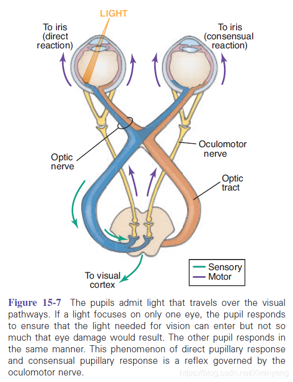
The pupillary light reflex causes pupils immediately to constrict when exposed to bright light. This can be seen as a direct reflex, in which constriction occurs in the eye exposed to the
light, or as an indirect or consensual reflex, in which exposure to light in one eye results in constriction of the pupil in the opposite eye (Fig. 15-7). These protective reflexes, mediated
by the oculomotor nerve, prevent damage to the delicate photoreceptors by excessive light.
Accommodation is a functional reflex allowing the eyes to focus on near objects. This is accomplished through movement of the ciliary muscles causing an increase in the curvature
of the lens. This change in shape of the lens is not visible.