包括的文章有:
一、Machine Learning Based Automatic Neovascularization Detection on Optic Disc Region.
二、Detection of neovascularization in retinal images using semi-supervised learning.
三、Proliferative diabetic retinopathy characterization based on the spatial organization of vascular junctions in fundus images
四、Automatic detection of neovascularization on optic disk region with feature extraction and support vector machine 5
五、Automated Detection of Neovascularization for Proliferative Diabetic Retinopathy Screening
六、Detection of neovascularisation using K-means clustering through registration of peripapillary OCT and fundus retinal images(使用K-means聚类通过配准周围OCT和眼底视网膜图像来检测新血管形成。)
七、Classification and detection of diabetic retinopathy using K-means algorithm(使用k-means算法对糖尿病视网膜病变做分类和检测)
一、Machine Learning Based Automatic Neovascularization Detection on Optic Disc Region
作者:Shuang Yu, Di Xiao and Yogesan Kanagasingam
数据:424幅视网膜图像,134幅有新生血管(NVD),290幅无新生血管(non-NVD)。
准确率:95.23%,特异性:96.30%,敏感性:92.90%
摘要:在本文中,介绍了眼底视网膜图像在视盘区域(NVD)中的新生血管形成的自动检测。 NV是增生性糖尿病视网膜病变(PDR)发病的指标,其特征在于视网膜中存在新血管。新生血管很脆弱,造成视力下降的风险很高。因此,不能低估NV的准确和及时检测的重要性。我们提出了一种用于NVD检测的自动图像处理过程,包括使用多级Gabor滤波的血管分割,血管形态特征和纹理特征的特征提取,以及支持向量机的图像分类。从每个NVD图像提取42个特征,并且特征选择过程进一步将最佳特征尺寸减小到18。所选特征在包含134个NVD和290个非NVD图像的424个视网膜图像上进行训练和测试。我们平均准确率为95.23%,特异度为96.30%,灵敏度为92.90%,AUC值为98.51%,随机选择试验组。
介绍:糖尿病视网膜病变是青年到中年人致盲和视觉损失的主要原因。糖尿病视网膜病变最严重的阶段是增生性阶段,其重要标志是新生血管的增殖。新生的血管很细,弯弯曲曲的,而且很脆弱,很容易断,高风险的导致严重的视力损失,甚至失明。
NVD 在视盘包括其附近一个视盘直径范围的视网膜出现新生血管,称为视盘新生血管。
NVE 在其他部分的视网膜新生血管称为视网膜新生血管。
方法:
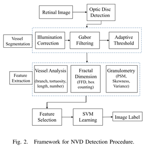
- 视盘探测:局部相位对称;
- 预处理:光照校正;非局部均匀滤波获得主血管;
- 血管分割:血管增强-多规模Gabor滤波器[26]在原图和主血管图中应用,得到所有血管和主血管的二值图像-两图相减-得到候选微血管。
- 特征提取
1)形态学特征:血管宽度,长度,曲率,加权长度和加权曲率;统计学特征,宽度,长度,曲率,加权长度和加权曲率的平均值,方差,偏态[30]。NVD:更小的宽度,更短的长度,更大的曲率,导致偏态值的变化。(15维)+血管数+血管分支数=17维*2=34维(一个在全血管图,一个在新生血管候选图)(34维)
曲率=血管像素长度/两端点欧氏距离;加权长度=血管像素长度*血管宽度;加权曲率=血管曲率*血管宽度。
2)纹理特征:新生血管密度更大,更无组织。提取fractal dimension[31]:分形维数,碎形维度:盒计数方法和傅里叶碎形维度方法。盒计数方法用于三幅二值图像(全部血管,粗血管,候选新生血管), 傅里叶碎形维度用于Gabor滤波后的原图Fig6(b),和出血管图Fig6(e)。粒度分析:模式谱的平均值,方差,偏态。(8维)
3)特征选择:选择,加权,最后选出最佳性能的特征子集。
E.支持向量机,k折交叉分类。
F. NVD数据:
MESSIDOR 4张
High-Resolution Fundus (HRF) 3张
DIARETDB0 11张
Kaggle DR database test 47 train 69
(134张)
Non-NVD数据
290张
(压缩为视盘半径为140)
结果和讨论:
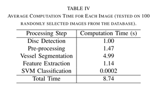
时间:8.74 second per image.
每类特征:
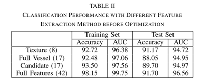
特征选择:
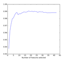
18个特征最高
In average, it achieves an accuracy of 95.23%, specificity of 96.30%, sensitivity of 92.90% and AUC of 98.51% on the test set, with the best performance of 95.29% accuracy, 95.16% specificity, 95.65% sensitivity and 99.27% AUC evaluated by the value of AUC.
对比:
精度:
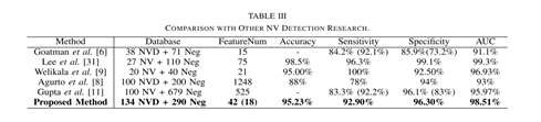
时间:
The accuracy is increased from 91.70% to 95.29% by the feature selection procedure。
二、Detection of neovascularization in retinal images using semi-supervised learning
作者:Appan K. Pujitha*, Gamalapati S. Jahnavi* and Jayanthi Sivaswamy
会议:ISBI 2017
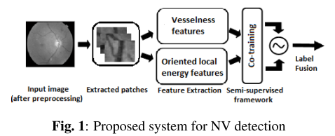
预处理:光照对比度标准化
特征提取:基于血管特征的Hessian矩阵。纹理特征。
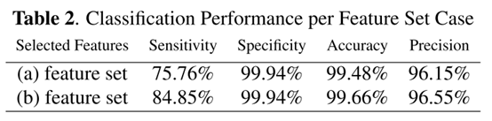
三、Proliferative diabetic retinopathy characterization based on the spatial organization of vascular junctions in fundus images
Some efforts have been placed towards automatic characterization of the NVs within the optic disc only (NVDs) [4,5]. NVDs are well contrasted against the background, owing to the fact that the optic disc is the brightest retinal structure. (However, the appearance of NVs, regardless of their site, constitute the transition to the proliferative stage, and it is thus important for the algorithms to be able to detect new vessels in all the available retinal sites.)
方法:
连接点中心探测:血管分割-预处理-张量投票(tensor voting)算法-连接点中心分离
空间分布测量:
结果:We apply a 10-fold feature selection process on two different feature sets: (a) including 39 features proposed in the literature, and (b) including 88 features, with 49 newly proposed.
四、Automatic detection of neovascularization on optic disk region with feature extraction and support vector machine
作者:Shuang Yu, Di Xiao and Yogesan Kanagasingam
根据AAO (America Academy of Ophthalmology )的推荐,根据疾病进展阶段,DR可分为轻度,中度和重度非增殖性DR(NPDR)和进一步增殖性DR(PDR)。
增殖期是最严重的状态。
视盘探测(局部相位对称)-血管分割(多维度Gabor 滤波器)-特征提取(统计学特征)
这篇文章是第一篇的会议版。
五、Automated Detection of Neovascularization for Proliferative Diabetic Retinopathy Screening
作者:Sohini Roychowdhury, Dara D. Koozekanani, and Keshab K. Parhi
会议:2016 38th Annual International Conference of the IEEE Engineering in Medicine and Biology Society (EMBC)
摘要:视盘,血管分割,特征提取(基于区域的特征),分类。
结果:NVD,sensitivity, specificity and accuracy,74%, 98.2%,87.6%, NVE:61%, 97.5%, 92.1%, PDR筛查,86.4% sensitivity and 76% specificity。
介绍:DR是发展中国家中青年致盲的主要原因。DR分为,非增殖性和增殖性。非增殖期的征兆有微动脉瘤,棉绒斑,硬性渗出和出血。取决于这些特征的出现,非增殖期DR 可分为轻度,中等,和重度。
本文贡献:1、分析比较了5个不同的图像滤波方法的重要性。高通滤波,形态学血管加强,梯度滤波,分水岭变换,Frangi滤波。高通滤波和形态学血管加强对于NVD/NVE检查比其他好。2、留一交叉验证法。选出最优特征子集。
方法:视盘分割(基于区域的分类)——血管分割(高通滤波)——
高通滤波:
形态学血管加强: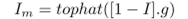
梯度滤波:
分水岭变换:
Frangi滤波: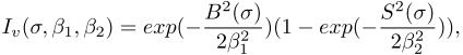


数据库:STARE:30 normal and 10 images with PDR. Local:All 17 images show varying severities of PDR.
While manifestations of NVD are visible as fine blood vessels in and around the major blood vessels in the OD region, instances of NVE appear as fine vessel-like abnormalities away from the OD region.
结果: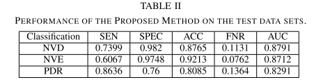
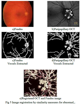
六、Detection of neovascularisation using K-means clustering through registration of peripapillary OCT and fundus retinal images(使用K-means聚类通过配准周围OCT和眼底视网膜图像来检测新血管形成。)
作者:N. Padmasini;R. Umamaheswari(印度人)
会议:2016 IEEE International Conference on Computational Intelligence and Computing Research (ICCIC)
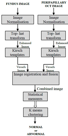
摘要:加强OCT图和眼底图,通过kirsch模板提取血管。两个图形通过基于配准的相似性测量融合。本文提出使用Kmeans聚类提取NV 特征来探测正常和不正常的血管。结果通过了所有实时样本,并得到医生的肯定。
Investigation of the blood vessels on the retinal images may have multiple applications from being pointers of different retinal diseases.
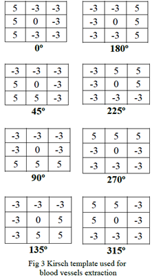
The Kirsch edge detection algorithm uses a single mask of size 3x3 and rotates it in 45 degree increments through all 8 directions.
高帽变换加强血管。
统计学参数:自相关,曲率,moment invariants(不变距),cluster prominence and cluster shade(簇突出和簇荫)。
分类器:K-means聚类

七、Classification and detection of diabetic retinopathy using K-means algorithm(使用k-means算法对糖尿病视网膜病变做分类和检测)
作者:S. B. ManojKumar,H. S. Sheshadri
会议:2016 International Conference on Electrical, Electronics, and Optimization Techniques (ICEEOT)
本文摘要:本文主要目的是一个识别背景型糖尿病视网膜病变和增值型糖尿病视网膜病变的框架。