OCELOT dataset
D = { ( x s , y s c , x l , y l t , c x , c y ) i } i = 1 N \mathcal{D} = \left\{\left(\mathbf{x}_s, \mathbf{y}_s^c, \mathbf{x}_l, \mathbf{y}_l^t, c_x, c_y\right)_i\right\}_{i=1}^{N} D={
(xs,ysc,xl,ylt,cx,cy)i}i=1N
x s ∈ R 1024 × 1024 × 3 \mathbf{x}_s\in\mathbb{R}^{1024\times 1024 \times 3} xs∈R1024×1024×3表示cell patch
y s C , ( y s C ) i ∈ R × R × { 1 , 2 } \mathbf{y}_s^C, \left(y_s^C\right)_i\in\mathbb{R}\times\mathbb{R}\times\left\{1, 2\right\} ysC,(ysC)i∈R×R×{
1,2}, 表示cell的标签,每个元素是一个三元组,1 Background Cell,2 Tumor Cell
x l ∈ R 1024 × 1024 × 3 \mathbf{x}_l\in\mathbb{R}^{1024\times 1024\times3} xl∈R1024×1024×3表示tissue patch
y l t ∈ { 1 , 2 , 255 } 1024 × 1024 \mathbf{y}_l^t\in\left\{1,2,255\right\}^{1024\times 1024} ylt∈{
1,2,255}1024×1024表示tissue的标签,1 Background,2 Cancer Area, 255 UNK (Unknown)
c x , c y c_x, c_y cx,cy是 x s \mathbf{x}_s xs的中心在 x l \mathbf{x}_l xl中的相对坐标
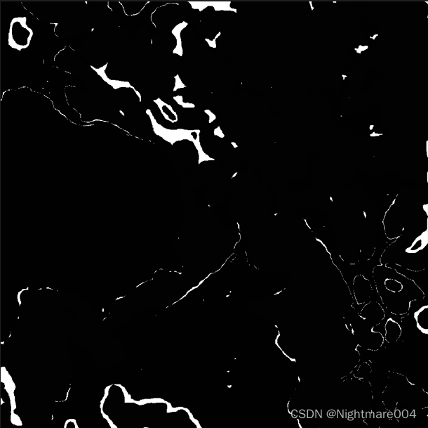
这是 y l t \mathbf{y}_l^t ylt,其中白色的部分是未知
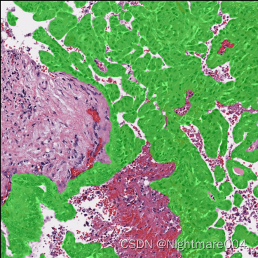
这是 x l \mathbf{x}_l xl,绿色部分是Cancer Area
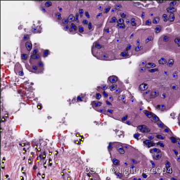
这是 x s \mathbf{x}_s xs,其中黄色的点为Background Cell,蓝色的点为Tumor Cell
配置
x s \mathbf{x}_s xs是 1024 × 1024 1024\times 1024 1024×1024,0.2MPP
x l \mathbf{x}_l xl是 4096 × 4096 4096\times 4096 4096×4096,0.2MPP, 随后4倍下采样到 1024 × 1024 1024\times 1024 1024×1024
BC:TC = 35.01%: 64.99%
BG:CA:UNK = 55.77%:40.17%:4.06%
训练:验证:测试=400:137:126
大约6:2:2
ps:
TC:BC = 0.36571231398085396: 0.6342876860191461
BG:CA:UNK = 0.5493605471304639: 0.4071568896148359: 0.043482563254700166
平均
train: BC:TC:all = 58.33:106.26.17:164.59
val: BC:TC:all = 61.31:119.07:180.38
test: BC:TC:all = 76.21:102.17:178.39
x s \mathbf{x}_s xs的原始尺寸:最小397, 最大1023, 平均801.8069381598793
x l \mathbf{x}_l xl的原始尺寸:最小1591, 最大4095, 平均3208.883861236802
原始尺寸 * mpp_x / resized_mpp_x = 1024
原始尺寸 * mpp_y / resized_mpp_y = 1024
能保证他们的原始尺寸是正方形(高=宽)
x l \mathbf{x}_l xl的原始尺寸: x s \mathbf{x}_s xs的原始尺寸的比例在4.00左右(即4或者4.00xxx)
x s \mathbf{x}_s xs在 x l \mathbf{x}_l xl的尺寸为256*256(因为原本就是在4096中的1024,现在4倍下采样,所以只剩下256*256)
实验
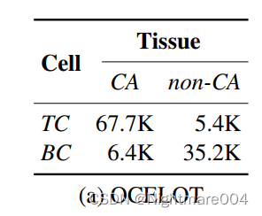
93%的TC在CA里
85%的BC在CA外面
因此细胞和组织一定有某种关系
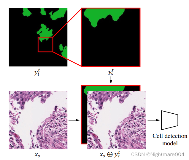
Tissue-label Leaking Model
将 y l t \mathbf{y}_l^t ylt对应于 x s \mathbf{x}_s xs的那一块切出来,然后resize到 1024 × 1024 1024\times 1024 1024×1024,表示为 y s t \mathbf{y}_s^t yst
随后将 x s \mathbf{x}_s xs和 y s t \mathbf{y}_s^t yst按通道concat,输入模型
在验证集和测试集上,F1分别提升7.69和9.76