Article Directory
1 Summary
For medical image segmentation, the author proposes a data enhancement strategy and one U-Net network structure by the end of the training structure able to use some of the images was better than the sliding window method (ordinary CNN) of effect. And, the network is very fast, the use of a 512x512 image segmentation ordinary GPU can be completed in less than one second.
2 Highlights
2.1 enhanced data
As the medical field image data set is relatively small, so how small amounts of data from the network structure of intensive training to get a good effect become a hot issue. And data employed herein means more enhanced by the image pan, rotate, adjust the image tone value, a random number of ways to enhance the elastic deformation data.
2.2 U-Net network structure
2.2.1 Crop
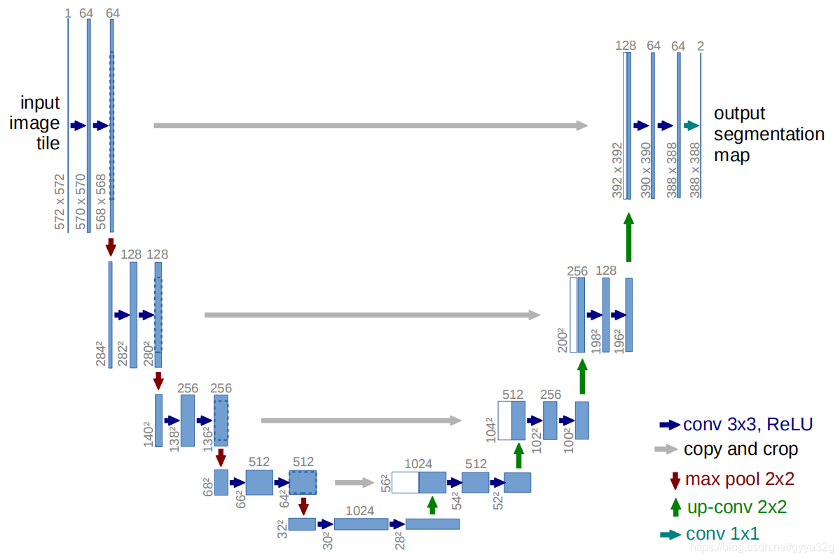
Overall analysis previously performed, first analyze local information - cut. From the overall network map, you can see the image gray arrow to the left of the image on the right is from big to small, such as through the gray arrow after 568x568 392x392 become the first line, this is a cutting process, you can see intermediate image to the left of the gray arrow with a box is the need to cut the size. In fact, from a macro perspective, the original input image size is 572x572, and the final output is 388x388. That input and output images simply do not match. First put the paper in the figure:
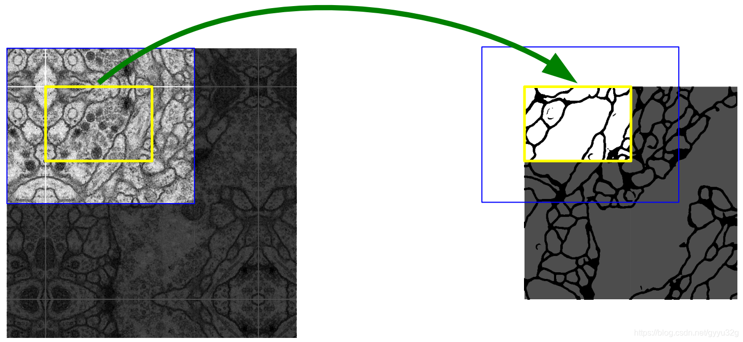
This article is to explain, if their prediction is required is left yellow area, then the beginning of the input image must first be extended to the area by counting the blue, the blue area can be said to be used to offset the cut of the network, without filling convolution to exactly predict the final output of the yellow region.
2.2.2 stitching feature
U-Net structure is a network structure of the FCN reference jump, and the general idea is the characteristic feature of FIG. FIG deep and shallow fusion, the network structure can be seen from the general view, in the right half of the U-Net will white and blue spliced feature maps, the white part of the sample from a substantially U-Net obtained green arrow, and then get shallow features gray arrows in FIG crop, characterized in splicing, unlike directly FCN through direct superposition of shallow and deep.
Analysis 2.2.3 U-Net overall structure
总框图中,最初地输入的图像为572x572,文章中采用的卷积方式是不填充的,所以每次卷积以后得到的图像会变小,得到568x568特征图,一方面进行一个池化操作,图像缩小;另一方面通过灰色箭头裁剪复制得到一个特征图。而第一步经过池化后的图像也执行同样的两个步骤:池化和裁剪复制。一直执行到图片大小变成28x28,进行上采样得到56x56特征图与上一次裁剪而得到的56x56图像进行拼接,然后进行卷积…上采样…拼接…卷积…上采样…拼接…卷积…最后经过一个1x1卷积得到388x388的图像输出。由于整个网络结构呈“U”形,所以为U-Net,而由于U形左边部分是特征图不断缩小的,文章称其为压缩路径,而右边部分的特征图不断地扩大,文章称其为扩展路径,这也算是一种编码-解码结构了。
3 效果
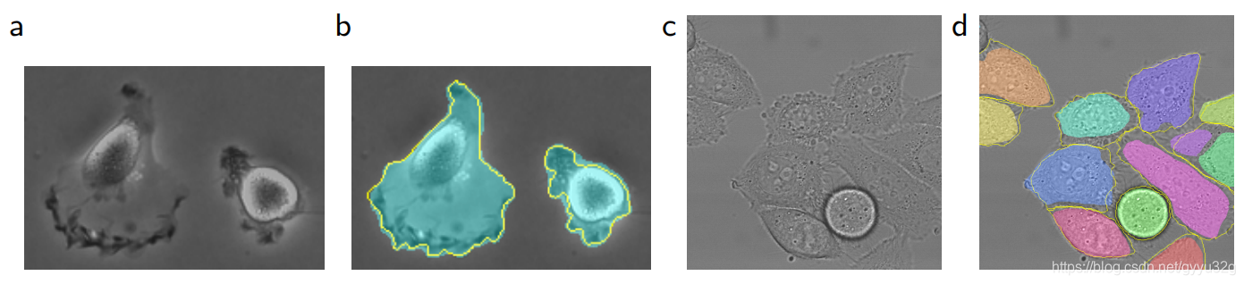
(a)为“PhC-U373” 数据集的输入,(b)的黄色边框为人工的分割结果,蓝色为网络分割结果;(c)为“DIC-HeLa” 数据集的输入,(d)的黄色边框为人工分割结果,彩色部分为网络分割结果。

上图为不同网络结构的IOU(交并比)的对比,可见u-net的效果表现最佳。
4 结论
本文提出的数据增强策略和U-Net网络结构确实能够使用非常少的训练样本而得到一个很好的医学上的分割效果,U-Net结构在医学图像分割领域有着重要的地位。
5 参考文献
(1)U-Net: Convolutional Networks for Biomedical Image Segmentation
(2)浅析U-Net是用来干嘛的
(3)深度学习图像分割——U-Net网络
